Veterinaria (Montevideo), 2026 62(226), 10.29155/VET.62.226.8
Revisión
The cytoskeleton as a functional axis of tumor aggressiveness in domestic animals: structural, mechanical, and translational integration
El citoesqueleto como eje funcional de la agresividad tumoral en animales domésticos: integración estructural, mecánica y traslacional
O citoesqueleto como eixo funcional da agressividade tumoral em animais domésticos: integração estrutural, mecânica e translacional
Sarai Guerrero-López1 https://orcid.org/0009-0008-2926-7450
Carlos Gerardo García-Tovar1 https://orcid.org/0000-0003-1890-8730
Samantha Jardon-Xicotencatl1 https://orcid.org/0000-0001-5259-8866
1Laboratorio 4, Morfología Veterinaria y Biología Celular, Unidad de Investigación Multidisciplinaria, Facultad de Estudios Superiores Cuautitlán, Universidad Nacional Autónoma de México, México. Corresponding author: doctora.jardon@cuautitlan.unam.mx
Cómo citar este artículo:
Guerrero-López, S., García-Tovar, C. G., & Jardon-Xicotencatl, S. (2026). The cytoskeleton as a functional axis of tumor aggressiveness in domestic animals: structural, mechanical, and translational integration. Veterinaria (Montevideo), 62(226). e20266222608. https://doi.org/10.29155/VET.62.226.7
Abstract
The cytoskeleton is a key regulatory system in tumor progression, integrating structural, biochemical, and mechanical signals that influence cellular behavior. This review critically analyzed evidence on cytoskeletal proteins and their remodeling as indicators of tumor aggressiveness in neoplasms of domestic animals, emphasizing dogs, cats, horses, and cattle. Literature from 2011 to 2025 was prioritized, with earlier seminal studies included for foundational or mechanistic context. Immunohistochemical, clinicopathological, and functional evidence indicates that actin reorganization, microtubule dynamics, altered intermediate filament expression, and E-cadherin-mediated adhesion are associated with invasion, proliferation, and cellular plasticity. Aberrant vimentin expression in epithelial neoplastic cells is associated with epithelial-mesenchymal plasticity, whereas its expression in mesenchymal neoplasms primarily reflects cellular lineage and should not independently indicate aggressiveness. However, veterinary evidence remains predominantly associative, with limited functional validation. Overall, the cytoskeleton represents a relevant functional axis of tumor aggressiveness with potential diagnostic and prognostic value, although further experimental validation is required.
Keywords: Cytoskeleton, Epithelial-mesenchymal plasticity, Tumor aggressiveness, Veterinary oncology, Domestic animals.
Resumen
El citoesqueleto es un sistema regulador clave en la progresión tumoral, que integra señales estructurales, bioquímicas y mecánicas que influyen en el comportamiento celular. Esta revisión analizó críticamente la evidencia sobre las proteínas del citoesqueleto y su remodelación como indicadores de agresividad tumoral en neoplasias de animales domésticos, con énfasis en perros, gatos, caballos y bovinos. Se priorizó la literatura publicada entre 2011 y 2025, incluyendo estudios seminales anteriores para proporcionar un contexto fundamental o mecanístico. La evidencia inmunohistoquímica, clínico-patológica y funcional indica que la reorganización de la actina, la dinámica de los microtúbulos, la expresión alterada de los filamentos intermedios y la adhesión mediada por E-cadherina se asocian con invasión, proliferación y plasticidad celular. La expresión aberrante de vimentina en células neoplásicas epiteliales se asocia con plasticidad epitelio-mesenquimal, mientras que su expresión en neoplasias mesenquimales refleja principalmente el linaje celular y no debe considerarse de manera independiente como indicadora de agresividad. Sin embargo, la evidencia veterinaria sigue siendo predominantemente asociativa, con una validación funcional limitada. En conjunto, el citoesqueleto representa un eje funcional relevante de la agresividad tumoral con potencial valor diagnóstico y pronóstico, aunque se requiere mayor validación experimental.
Palabras clave: Citoesqueleto, Plasticidad epitelio-mesenquimal, Agresividad tumoral, Oncología veterinaria, Animales domésticos.
Resumo
O citoesqueleto é um sistema regulador chave na progressão tumoral, integrando sinais estruturais, bioquímicos e mecânicos que influenciam o comportamento celular. Esta revisão analisou criticamente as evidências sobre as proteínas do citoesqueleto e sua remodelação como indicadores de agressividade tumoral em neoplasias de animais domésticos, com ênfase em cães, gatos, cavalos e bovinos. Foi priorizada a literatura publicada entre 2011 e 2025, incluindo estudos seminais anteriores para fornecer um contexto fundamental ou mecanístico. Evidências imuno-histoquímicas, clinicopatológicas e funcionais indicam que a reorganização da actina, a dinâmica dos microtúbulos, a expressão alterada dos filamentos intermediários e a adesão mediada pela E-caderina estão associadas à invasão, proliferação e plasticidade celular. A expressão aberrante de vimentina em células neoplásicas epiteliais está associada à plasticidade epitélio-mesenquimal, enquanto sua expressão em neoplasias mesenquimais reflete principalmente a linhagem celular e não deve ser considerada, de forma independente, um indicador de agressividade. No entanto, as evidências veterinárias permanecem predominantemente associativas, com validação funcional limitada. De modo geral, o citoesqueleto representa um eixo funcional relevante da agressividade tumoral, com potencial valor diagnóstico e prognóstico, embora seja necessária maior validação experimental.
Palavras-chave: Citoesqueleto, Plasticidade epitélio-mesenquimal, Agressividade tumoral, Oncologia veterinária, Animais domésticos.
Fecha de recibido: 06/05/2026
Fecha de aceptado: 02/10/2026
Introduction
Cancer is defined as a dynamic process driven by the accumulation of genomic and epigenetic alterations that transform normal cells into cells with exceptional biological capabilities, known as the “hallmarks of cancer”. These capabilities, recently updated to include dimensions such as metabolic reprogramming and immune evasion, include uncontrolled proliferation, evasion of growth suppressors, and activation of mechanisms of invasion and metastasis (Hanahan, 2022; Vogelstein et al., 2013). In veterinary medicine, neoplasms represent one of the leading causes of morbidity and mortality in companion animals, establishing tumors in dogs and cats as highly relevant biological models for comparative oncology (Lane, 2018; Paoloni & Khanna, 2007). This discipline recognizes that cancer shares fundamental signaling pathways across species, which is essential for the development of novel diagnostic and therapeutic strategies (Vail et al., 2020).
Within this biological context, the cytoskeleton emerges as a central regulatory system that integrates cellular architecture, signaling, and mechanical responses, allowing cells to maintain their shape and interact with the microenvironment. During malignant transformation, this system becomes a key effector of cellular movement and invasion. Reorganization of actin filaments, microtubules, and intermediate filaments enables tumor cells to withstand intracellular stress and adapt to the mechanical conditions of the neoplastic environment (Fife et al., 2014; Ong et al., 2020). This structural plasticity facilitates directed cell migration by promoting detachment from the primary tumor and movement through the extracellular matrix (Bravo-Cordero et al., 2012).
Studies of the tumor microenvironment have further revealed that cancer progression is not solely a genetic and molecular phenomenon, but also a mechanical one. The cytoskeleton acts as a mechanotransduction system that senses physical cues from the environment, such as tissue stiffness, and converts them into cellular responses that may promote invasion and tumor progression (Li & Wang, 2020). Within this process, epithelial-mesenchymal plasticity is particularly relevant because it involves coordinated changes in cell adhesion, cytoskeletal organization, and signaling pathways that enable the acquisition of migratory and invasive phenotypes.
Therefore, the objective of this review is to evaluate the available evidence regarding the role of cytoskeletal proteins and their remodeling as indicators of aggressiveness in canine, feline, equine, and bovine neoplasms, while integrating a conceptual synthesis of cellular plasticity mechanisms that link structural architecture with tumor behavior. By focusing on species of relevance to comparative oncology, this review seeks to establish a critical framework that distinguishes the physiological expression of cytoskeletal proteins from alterations associated with neoplastic transformation, while also differentiating clinicopathological findings from functional mechanisms described in experimental models.
For this critical review, a literature search was conducted in the PubMed and ScienceDirect databases, primarily focusing on scientific publications published between 2011 and 2025, while earlier seminal studies were included when necessary to provide foundational or mechanistic context. Search terms were related to the cytoskeleton, epithelial-mesenchymal plasticity, tumor aggressiveness, and comparative oncology. The analysis focused on canine, feline, equine, and bovine neoplasms, prioritizing studies that evaluated cytoskeletal proteins, adhesion molecules, and signaling pathways associated with cytoskeletal remodeling and tumor progression. Immunohistochemical, clinicopathological, and experimental studies were integrated, distinguishing the physiological expression of lineage-associated proteins from their aberrant expression or overexpression in neoplastic cells. Findings directly demonstrated in veterinary neoplasms were also distinguished from functional mechanisms derived from experimental models or human oncology.
Normal cytoskeletal organization and cell-type distribution
The cytoskeleton is an integrated structural network composed of actin filaments, microtubules, and intermediate filaments, which collectively maintain cell shape, mechanical stability, intracellular organization, and motility. Actin filaments are composed of polymerized actin and participate in cortical organization, cell adhesion, and the generation of forces required for cellular movement. Microtubules are formed by α- and β-tubulin heterodimers and contribute to cell polarity, intracellular transport, and mitotic spindle organization. Unlike these components, intermediate filaments exhibit a more pronounced cell-type-specific distribution and therefore provide useful information regarding cellular lineage and differentiation (Akhmanova & Steinmetz, 2015; Herrmann & Aebi, 2016; Ong et al., 2020).
Under physiological conditions, cytokeratins are predominantly expressed in epithelial cells, whereas vimentin is characteristic of cells of mesenchymal origin. Other intermediate filament proteins show similarly defined distributions, including desmin in muscle cells, glial fibrillary acidic protein (GFAP) in glial cells, and neurofilaments in neurons (Herrmann & Aebi, 2016; Ong et al., 2020). These normal expression patterns provide a necessary reference for interpreting cytoskeletal findings in neoplasms. Therefore, the presence of a specific intermediate filament in a tumor does not necessarily represent aberrant expression, because neoplastic cells may retain the cytoskeletal profile of their cell of origin. Conversely, the acquisition or altered expression of lineage-associated proteins may reflect changes in cellular differentiation and plasticity during tumor progression (Ong et al., 2020).
This distinction is particularly relevant to the interpretation of vimentin throughout this review. Its expression is physiological in mesenchymal cells, including fibroblasts, whereas its expression in epithelial tumor cells may indicate cytoskeletal reprogramming associated with epithelial-mesenchymal plasticity (Danielsson et al., 2018; Mendez et al., 2010; Usman et al., 2021). Accordingly, the cytoskeletal alterations discussed in the following sections are interpreted according to normal cellular lineage, distinguishing retained physiological expression from aberrant expression or overexpression associated with neoplastic progression. The principal components and their normal cell-type distribution are summarized in figure 1.
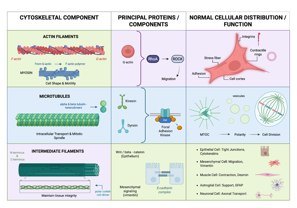
Figure 1: Normal cytoskeletal organization and cell-type distribution. Schematic representation of the three major cytoskeletal systems and their principal physiological distribution. Actin filaments and microtubules contribute to cell architecture, motility, polarity, intracellular transport, and cell division, whereas intermediate filaments exhibit greater cell-type specificity. Cytokeratins are predominantly associated with epithelial cells, vimentin with mesenchymal cells, desmin with muscle cells, GFAP with glial cells, and neurofilaments with neurons.
Cytoskeletal alterations and tumor progression
Actin Filaments
Tumor progression toward metastatic stages depends closely on the ability of neoplastic cells to reorganize their actin filaments, transforming a relatively static structure into a dynamic machinery for movement and invasion. According to Yamaguchi and Condeelis (2007), regulation of the actin cytoskeleton enables the formation of specialized protrusions such as lamellipodia and filopodia, which function as sensory and propulsive structures that guide cells through the tumor microenvironment. However, migration is not the only challenge; for true invasion to occur, tumor cells must overcome the physical barriers imposed by the extracellular matrix. Invadopodia are actin-rich structures associated with degradative enzymes that function as specialized invasive structures, allowing tumor cells to penetrate adjacent tissues and gain access to circulatory pathways. Thus, actin dynamics not only confer motility but also influence the directionality and efficiency with which cancer cells colonize distant sites within the organism (Bravo-Cordero et al., 2012).
Microtubules
In addition to serving as structural scaffolds, microtubules are dynamic components essential for both the proliferation and adaptation of cancer cells. The organization of these filaments depends on precise regulation at their ends, where the balance between assembly and disassembly allows cells to maintain polarity and efficiently organize intracellular organelle transport, processes that are frequently altered in cancer to favor migration (Akhmanova & Steinmetz, 2015). This dynamic instability becomes particularly important during mitosis, when microtubules form the mitotic spindle. Alterations in microtubule dynamics may promote defects in cell division and contribute to tumor progression. Thus, microtubules not only provide physical support but also participate in processes essential for tumor cell proliferation, survival, and motility (Ritter & Kreis, 2022).
Intermediate Filaments
Intermediate filaments, whose structure and assembly provide cells with unique mechanical stability (Herrmann & Aebi, 2016), play a prominent role in tumor progression, with vimentin representing one of the most relevant components in the context of tumor plasticity. During epithelial-mesenchymal plasticity (EMP), vimentin expression is associated with changes in cell morphology, increased motility, and altered cell adhesion (Mendez et al., 2010). Vimentin is not merely a lineage marker; it also participates in complex processes within the tumor microenvironment, including invasion, immune evasion, and cellular metabolism (Parvanian & Eriksson, 2025; Usman et al., 2021).
However, interpretation of vimentin expression depends on cellular lineage. Its presence is physiological in cells of mesenchymal origin and therefore does not, by itself, constitute an indicator of neoplastic transformation or aggressiveness. In contrast, its expression in neoplastic cells of epithelial origin may represent part of the cytoskeletal reprogramming associated with epithelial-mesenchymal plasticity. Given its functional diversity in health and disease (Danielsson et al., 2018), the relevance of vimentin as a biomarker should be interpreted according to cell type, expression pattern, and neoplastic context (Satelli & Li, 2011).
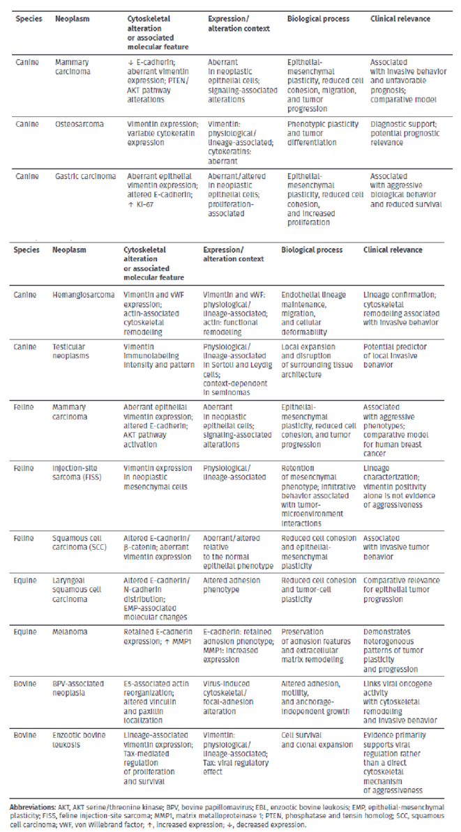
Collectively, the three cytoskeletal systems do not operate independently but rather as an integrated functional network that coordinates motility, proliferation, and adaptation to mechanical stress. Actin drives migration and invasion, microtubules maintain polarity and support cell division, and intermediate filaments provide mechanical stability, while their reorganization participates in EMP. This functional convergence constitutes the structural basis of tumor plasticity, a phenomenon observed across multiple veterinary neoplasms discussed in the following sections (Table 1).
Table 1. Cytoskeletal alterations and associated molecular features related to tumor aggressiveness in domestic animal neoplasms
Epithelial-mesenchymal plasticity (EMP) and cytoskeletal dynamics
Modern carcinogenesis is understood as a dynamic process involving epithelial-mesenchymal plasticity (EMP). This concept extends beyond the traditional view of epithelial-mesenchymal transition (EMT) as a binary switch and instead recognizes a continuous spectrum in which tumor cells may transition through hybrid epithelial/mesenchymal (E/M) states (Sahoo et al., 2022; Usman et al., 2021). In these intermediate states, cells retain epithelial markers while acquiring mesenchymal properties, thereby gaining enhanced plasticity associated with metastasis, immune evasion, and therapeutic resistance (Sahoo et al., 2022; Usman et al., 2021).
Molecular Regulation and Signaling
This process is orchestrated by a complex network of microenvironmental inducers, including transforming growth factor beta (TGF-β), Wnt/β-catenin signaling, and hypoxia (Li & Wang, 2020; Usman et al., 2021). These signals activate central transcriptional regulators such as SNAIL, TWIST, and ZEB1/2, which repress genes involved in cell cohesion, particularly E-cadherin (Hanahan, 2022; Usman et al., 2021). Loss of membrane-associated E-cadherin, a calcium-dependent adhesion protein essential for tissue integrity, allows cells to lose apicobasal polarity and detach from the primary tumor mass (Hanahan, 2022; Pimenta et al., 2023; Usman et al., 2021).
Cytoskeletal Reorganization during EMP
During EMP, coordinated cytoskeletal reorganization occurs: cells modify the organization of their cytokeratin filaments and assemble a dynamic vimentin network, which contributes to nuclear protection during migration and is associated with survival pathways such as PI3K/AKT/mTOR (Danielsson et al., 2018; Mendez et al., 2010; Usman et al., 2021). In parallel, actin microfilaments reorganize to form motile protrusions such as lamellipodia and invasive structures known as invadopodia (Bravo-Cordero et al., 2012; Yamaguchi & Condeelis, 2007).
Role of Matrix Metalloproteinases (MMPs)
Although matrix metalloproteinases (MMPs) are not structural components of the cytoskeleton, they are included in this review because of their functional relationship with invasion processes associated with cytoskeletal remodeling. Actin reorganization in structures such as invadopodia is associated with the localization of degradative enzymes that act on the extracellular matrix, thereby facilitating tumor invasion. In this context, MMP1, MMP2, and MMP9 are considered functional effectors associated with extracellular matrix degradation rather than components of the cytoskeleton itself (Brodesser et al., 2024; Parvanian & Eriksson, 2025).
Alterations of the cytoskeleton in specific tumors
Canines
Dogs are considered fundamental models in comparative oncology because they spontaneously develop tumors that share biological, genomic, and therapeutic response characteristics with human cancers (Gardner et al., 2016). In this species, the cytoskeleton not only provides structural support, but its remodeling has also been identified as a functional axis associated with biological aggressiveness across several cancer types (Li & Wang, 2020).
Canine Mammary Carcinoma
Canine mammary carcinoma (CMC) is characterized by marked biological heterogeneity, with tumor aggressiveness associated with the induction of epithelial-mesenchymal plasticity (EMP) (Gardner et al., 2016; Nosalova et al., 2024). A central event in this progression is the disruption of physiological cell-cell cohesion, normally mediated by membrane-associated E-cadherin. In invasive carcinomas, such as the micropapillary subtype, this process is associated with activation of the transcriptional repressor SNAIL, which binds to the promoter region of the CDH1 gene and suppresses E-cadherin expression (Flores et al., 2022; Gamba et al., 2015). Loss of adherens junctions facilitates disruption of epithelial organization and infiltration of surrounding tissues, contributing to the transition from an in situ to an invasive phenotype (Gamba et al., 2015).
From a structural perspective, malignant transformation is associated with aberrant vimentin expression in luminal epithelial cells, which under physiological conditions predominantly express cytokeratins (Fife et al., 2014; Flores et al., 2022; Sammarco et al., 2023). This alteration in intermediate filament expression contributes to increased mechanical resilience and occurs within a signaling context involving the PTEN/AKT pathway. Immunohistochemical studies have shown loss of PTEN expression in a subset of carcinomas, accompanied by increased AKT activation (p-AKT). Activation of this pathway is associated with processes related to cytoskeletal remodeling, migration, and tumor progression and has been linked to an unfavorable clinical prognosis (Asproni et al., 2021).
Canine Gastric Carcinoma
Canine gastric carcinoma (CGC) is a low-incidence neoplasm with markedly aggressive biological behavior and a high frequency of metastatic disease at diagnosis (Flores et al., 2022; Li & Wang, 2020). Within this context, structural alterations associated with malignancy include aberrant vimentin expression in neoplastic cells of epithelial origin (Gardner et al., 2016; Hardas et al., 2021). It is important to distinguish this finding from the physiological expression of vimentin in the tumor-associated stroma, where fibroblasts normally express this intermediate filament. In contrast, its acquisition by luminal epithelial tumor cells is consistent with epithelial-mesenchymal plasticity (EMP). This process is accompanied by alterations in E-cadherin-mediated adherens junctions, including reduced membranous expression and abnormal cytoplasmic localization, which weaken tissue architecture and facilitate infiltration into deeper tissues (Hardas et al., 2021).
The biological aggressiveness of CGC is further associated with the relationship between cytoskeletal remodeling and cell proliferation. The combination of epithelial vimentin expression and a high Ki-67 proliferation index identifies tumors with a more aggressive biological phenotype and has been associated with reduced patient survival (Flores et al., 2022). CD44 expression in neoplastic epithelial cells is also relevant in this context because, although CD44 is not a cytoskeletal component, it functions as a signaling platform or coreceptor associated with cell motility and survival (Hardas et al., 2021). Together with cytoskeletal alterations, CD44 expression may contribute to cellular interactions involved in metastatic dissemination (Hardas et al., 2021).
Canine Osteosarcoma
Osteosarcoma is the most common primary bone neoplasm in dogs and is characterized by marked microscopic heterogeneity and locally aggressive behavior. This histological diversity represents a diagnostic challenge that may be addressed through the evaluation of cytoskeletal proteins. Because osteosarcoma is a tumor of mesenchymal lineage, vimentin expression is expected as part of its physiological lineage-associated phenotype. However, aberrant expression of several cytokeratins, proteins normally associated with epithelial cells, has been reported together with vimentin in different histological subtypes. This variability in cytoskeletal composition reflects phenotypic plasticity and differentiation within the tumor (Gardner et al., 2016; Nagamine et al., 2015).
From a signaling perspective, cytoskeletal remodeling may be associated with pathways such as PI3K/AKT. Alterations involving the tumor suppressor PTEN and activation of AKT contribute to cell survival and anchorage-independent growth (Asproni et al., 2021; Bird & Smith, 2025). From a prognostic standpoint, the intensity and pattern of vimentin expression have been investigated in association with clinical behavior. These findings must be interpreted in the context of its physiological expression in mesenchymal cells rather than as evidence of aggressiveness based solely on marker positivity. Bone matrix-associated markers such as osteonectin (SPARC) and osteocalcin have also been evaluated in relation to tumor differentiation and prognosis (Amaral et al., 2018; Hanahan, 2022).
Canine Hemangiosarcoma
Canine hemangiosarcoma (HSA) is a malignant neoplasm of vascular endothelial origin characterized by high invasive and metastatic potential. Because of its marked morphological variability, immunohistochemical markers are important for lineage identification. In this context, it is essential to distinguish that vimentin and von Willebrand factor (vWF) expression is associated with endothelial lineage rather than representing, by itself, an acquired marker of tumor aggressiveness. Unlike epithelial carcinomas, in which vimentin expression may reflect an acquired mesenchymal phenotype, its presence in HSA is consistent with the normal phenotype of endothelial cells and may persist after neoplastic transformation (Li & Wang, 2020; Sabattini & Bettini, 2009).
Beyond lineage identification, the aggressive behavior of HSA is associated with functional changes that support cell migration and deformability. Cytoskeletal remodeling, particularly involving actin, contributes to the mechanical properties required for tumor cell movement and invasion (Bravo-Cordero et al., 2012; Fife et al., 2014). These structural changes should therefore be distinguished from the constitutive expression of lineage markers such as vimentin and vWF.
Signaling pathways associated with cell survival and motility, including PI3K/AKT/mTOR, have also been implicated in neoplastic progression. However, these signaling molecules are not structural components of the cytoskeleton; rather, they provide regulatory context for processes that may influence cytoskeletal organization and tumor behavior (Asproni et al., 2021). Likewise, molecules such as CD44 may participate in signaling processes related to adhesion and motility and should be interpreted as functionally associated factors rather than cytoskeletal proteins (Asproni et al., 2021; Sabattini & Bettini, 2009).
Testicular Neoplasms
In canine testicular neoplasms, interpretation of vimentin expression requires consideration of the normal cell lineage. In Sertoli and Leydig cell tumors, vimentin expression is physiological because these cells normally express this intermediate filament as part of their cytoskeletal phenotype (de Barros et al., 2025). Therefore, tumor aggressiveness should not be inferred from the mere presence of vimentin. Instead, differences in staining intensity and expression pattern have been evaluated in relation to local invasion and tumor expansion. Stronger vimentin immunolabeling, particularly in diffuse or expansile growth patterns, has been associated with more extensive disruption of surrounding tissue and may therefore have predictive value for local invasive behavior (de Barros et al., 2025).
In seminomas, interpretation is more complex because vimentin expression may differ according to the neoplastic phenotype and should not automatically be considered equivalent to epithelial-mesenchymal plasticity. Accordingly, its presence must be evaluated in relation to the normal phenotype of germ cells, tumor architecture, and the expression pattern reported in the original study. In this context, intense vimentin immunolabeling has been associated with more extensive local growth and disruption of the surrounding testicular architecture, supporting its potential value as an indicator of local biological behavior rather than as direct evidence of a mechanistic EMP process (de Barros et al., 2025).
Felines
In feline medicine, the study of the cytoskeleton has gained particular relevance in neoplasms characterized by aggressive and infiltrative behavior (Harris et al., 2019; Zappulli et al., 2015). As in dogs, alterations in the organization and expression of cytoskeletal proteins may accompany tumor progression and changes in invasive capacity. However, their interpretation requires consideration of the cellular lineage of each neoplasm, because the expression of specific cytoskeletal components may represent either retention of a physiological phenotype or aberrant expression associated with tumor plasticity (Sammarco et al., 2023).
Feline Mammary Carcinoma
Feline mammary carcinoma (FMC) is characterized by aggressive biological behavior, with a high proportion of malignant tumors and frequent development of metastases (Sammarco et al., 2023; Zappulli et al., 2015). Among the signaling pathways associated with its progression is the PI3K/AKT/mTOR pathway, whose activation promotes cell survival and inhibition of apoptosis (Asproni et al., 2021). In this context, loss of expression of the tumor suppressor PTEN, described in a substantial proportion of these tumors, may contribute to dysregulation of AKT signaling (Asproni et al., 2021; Maniscalco et al., 2012).
From a cytoskeletal perspective, epithelial-mesenchymal plasticity (EMP) provides a framework for interpreting the changes observed in neoplastic cells. Whereas luminal epithelial cells physiologically maintain a phenotype characterized by cytokeratin expression and membranous E-cadherin, vimentin expression has been described in neoplastic epithelial cells in certain FMCs, particularly in more aggressive phenotypes (Sammarco et al., 2023). In this context, vimentin expression should be considered aberrant relative to the luminal epithelial phenotype and may be interpreted as evidence of cytoskeletal reprogramming associated with EMP. In parallel, alterations in E-cadherin expression and localization may weaken intercellular cohesion and favor an invasive phenotype (Caliari et al., 2014; Sammarco et al., 2023).
Matrix metalloproteinases, including MMP2 and MMP9, are not components of the cytoskeleton and should therefore be considered functional effectors associated with extracellular matrix degradation. Their activity may facilitate invasion when it converges with changes in cell adhesion and cytoskeletal organization that accompany tumor plasticity (Parvanian & Eriksson, 2025; Usman et al., 2021).
Feline Injection-Site Sarcoma (FISS)
Feline injection-site sarcoma (FISS) is a mesenchymal neoplasm characterized by locally infiltrative behavior and an association with persistent inflammatory processes at injection sites (de Cecco et al., 2021; Hartmann et al., 2015). In this tumor, the interpretation of vimentin expression requires particular caution. Because of its mesenchymal origin, vimentin expression is consistent with the physiological phenotype of fibroblasts and other mesenchymal cells; therefore, its presence in neoplastic cells does not, by itself, constitute aberrant expression or evidence of EMP.
Consequently, unlike the situation observed in carcinomas of epithelial origin, vimentin positivity in FISS should primarily be interpreted as retention of a lineage-associated marker. The marked infiltrative capacity of this neoplasm cannot be attributed solely to the presence of vimentin but should instead be understood in the context of the functional alterations accompanying neoplastic transformation and the interactions between tumor cells, the inflammatory microenvironment, and the extracellular matrix (de Cecco et al., 2021; Hartmann et al., 2015). This distinction is particularly important to avoid interpreting a protein physiologically expressed by fibroblasts as an acquired marker of tumor aggressiveness.
Feline Squamous Cell Carcinoma (SCC)
Feline squamous cell carcinoma (SCC) is characterized by locally invasive behavior and the ability to infiltrate adjacent tissues (Harris et al., 2019). Invasive progression is associated with alterations in cell adhesion mechanisms, particularly those involving the E-cadherin/β-catenin complex. Reduced or altered membranous expression of these proteins weakens cohesion between epithelial cells and may favor the acquisition of an invasive phenotype. Alterations in β-catenin localization are also relevant because of its dual role as a component of adherens junctions and a mediator of the Wnt signaling pathway (Sanz Ressel et al., 2023).
From a cytoskeletal perspective, vimentin expression in neoplastic squamous cells represents an alteration relative to the normal epithelial phenotype, in which cytokeratins predominate. Therefore, unlike the situation observed in mesenchymal neoplasms such as FISS, acquisition of vimentin expression by neoplastic epithelial cells may be interpreted in the context of epithelial-mesenchymal plasticity (Sanz Ressel et al., 2023). The coexistence of epithelial and mesenchymal features is consistent with partial or hybrid EMP states, in which tumor cells retain certain components of epithelial adhesion while acquiring structural characteristics associated with increased motility and invasion (Harris et al., 2019; Sanz Ressel et al., 2023).
Equines
In horses, the study of cytoskeletal alterations and molecules associated with cell adhesion and plasticity has gained increasing interest in neoplasms with locally aggressive behavior, such as squamous cell carcinoma, as well as in tumors with distinctive biological characteristics, such as melanoma in gray horses (Strohmayer et al., 2022). In this context, horses represent a relevant model for evaluating the relationship among cellular plasticity, adhesion, and neoplastic progression, including the potential influence of genetic factors and, in certain epithelial neoplasms, viral agents such as equine papillomavirus type 2 (EcPV2) (Strohmayer et al., 2022).
Equine Laryngeal Squamous Cell Carcinoma
Equine squamous cell carcinoma (SCC) is an epithelial neoplasm that may exhibit locally invasive behavior. In its laryngeal presentation, alterations consistent with epithelial-mesenchymal plasticity have been described, including changes in the expression and localization of cell adhesion molecules (Armando et al., 2020). Whereas normal squamous epithelium physiologically maintains an organization based on cytokeratins and E-cadherin-mediated adherens junctions, neoplastic cells may exhibit alterations in the distribution of E-cadherin and N-cadherin. These changes are consistent with altered cell cohesion and the acquisition of features associated with a more invasive phenotype (Armando et al., 2020; Strohmayer et al., 2022).
In this context, EcPV2 infection has been associated with certain equine SCCs, and the expression of viral proteins may contribute to alterations in cellular mechanisms involved in neoplastic transformation (Armando et al., 2020; Strohmayer et al., 2022). In addition, signaling pathways and transcriptional regulators associated with epithelial-mesenchymal plasticity, including AKT, MAPK, TWIST, ZEB, and SNAIL, provide a mechanistic framework for interpreting the changes in cell adhesion and organization observed during tumor progression. However, these mechanisms should be distinguished from structural components of the cytoskeleton, and their specific involvement should be interpreted according to the evidence available for equine SCC (Armando et al., 2020; Hanahan, 2022).
Alterations in cell adhesion and plasticity may functionally converge with the reorganization of the actin cytoskeleton required for cell migration and invasion. Similarly, CD44 and CD271 are not cytoskeletal components but rather molecules associated with cellular phenotypes and signaling processes; therefore, their expression should be interpreted as complementary evidence of tumor heterogeneity and plasticity rather than as a direct structural alteration of the cytoskeleton (Armando et al., 2020; Strohmayer et al., 2022).
Equine Melanoma
Melanoma is a common neoplasm in gray horses and exhibits distinctive biological characteristics associated with genetic factors, including a duplication affecting the STX17 region (Brodesser et al., 2024). Many of these tumors may exhibit a prolonged clinical course before developing more aggressive behavior. In this context, adhesion molecules provide relevant information regarding the tumor phenotype. Although E-cadherin is not a component of the cytoskeleton, it participates in adherens junctions and establishes a functional connection between intercellular adhesion and cytoskeletal organization. Its expression has been described in a substantial proportion of equine melanomas, including some tumors with histological features of malignancy (Brodesser et al., 2024; Pimenta et al., 2023).
This finding should be interpreted with caution, because retention of E-cadherin does not, by itself, demonstrate benign biological behavior or allow the absence of invasion or metastasis to be directly attributed to its expression. Rather, its retention indicates that some equine melanomas may preserve features of cell adhesion during tumor progression, demonstrating that changes associated with tumor plasticity do not necessarily follow a uniform pattern (Pimenta et al., 2023).
The transcription factor MITF also participates in the regulation of the melanocytic phenotype and cellular plasticity; however, it is not a component of the cytoskeleton and should therefore be considered a molecular regulator within this context (Brodesser et al., 2024; Li & Wang, 2020). Similarly, matrix metalloproteinases (MMPs) are extracellular effectors rather than cytoskeletal proteins. Their inclusion is relevant because of their role in extracellular matrix degradation and, consequently, their functional relationship with cellular mechanisms of invasion. Among these enzymes, increased MMP1 expression has been described in equine melanoma tissue, supporting the involvement of extracellular matrix remodeling in tumor progression (Brodesser et al., 2024).
Bovines
Bovine Tumors
The relevance of the cytoskeleton in bovine oncology is particularly evident in neoplastic processes associated with infectious agents, including bovine papillomavirus (BPV) and bovine leukemia virus (BLV). In contrast to the predominantly spontaneous tumors described in companion animals, virus-associated bovine neoplasms provide an opportunity to examine how viral proteins can interfere with host-cell signaling and structural organization, thereby contributing to cellular transformation, clonal expansion, and tissue invasion. These tumors therefore provide a useful framework for examining the relationship between viral infection, cytoskeletal remodeling, and neoplastic progression.
Bovine Papillomavirus-Associated Neoplasia (BPV)
In bovine papillomavirus-associated neoplasia, the E5 oncoprotein of BPV-1 and BPV-4 has been associated with alterations in cytoskeletal organization during cellular transformation. Under physiological conditions, actin microfilaments contribute to cortical tension, cell shape, and the organization of focal adhesions. Expression of E5 induces marked reorganization of the actin cytoskeleton, including shortening of actin filaments and disruption of their normal extension toward the plasma membrane. These structural changes are accompanied by altered localization of focal adhesion-associated proteins, including vinculin and paxillin, from peripheral adhesion sites toward intracellular compartments (Borzacchiello & Roperto, 2008; Tsirimonaki et al., 2006).
Mechanistically, these alterations have been associated with changes in c-Src tyrosine kinase signaling, linking viral transformation to cytoskeletal remodeling, altered cell adhesion, and anchorage-independent growth. Importantly, vinculin and paxillin are not structural filaments of the cytoskeleton but focal adhesion-associated proteins that functionally connect the actin cytoskeleton to adhesion complexes and intracellular signaling. Although E5 expression from both BPV types has been associated with increased cell motility, experimental evidence indicates that BPV-1 E5 confers a greater invasive capacity in extracellular matrix models, supporting a functional relationship between viral oncogene expression, cytoskeletal remodeling, and invasive behavior (Borzacchiello & Roperto, 2008; Tsirimonaki et al., 2006).
Enzootic Bovine Leukosis
Enzootic bovine leukosis (EBL) is a lymphoproliferative disease caused by bovine leukemia virus (BLV), in which neoplastic transformation primarily involves B lymphocytes. In normal B lymphocytes, vimentin constitutes a major intermediate filament component and therefore represents physiological lineage-associated expression rather than a neoplasm-specific marker. Consequently, its presence in BLV-associated lymphoid cells should not itself be interpreted as evidence of cytoskeletal reprogramming.
BLV-induced transformation involves the viral Tax protein, a regulatory oncoprotein that alters cellular transcription and signaling pathways involved in proliferation and survival (Gillet et al., 2007). Although cytoskeletal alterations and syncytium formation may be observed in experimental systems involving BLV infection, these phenomena should be distinguished from the mechanisms responsible for naturally occurring EBL and lymphosarcoma. In particular, syncytium formation does not by itself demonstrate that cytoskeletal remodeling is the primary mechanism underlying neoplastic progression in vivo.
Therefore, in BLV-associated neoplasms, the available evidence more strongly supports a role for viral regulation of cell proliferation, survival, and clonal expansion than a direct cytoskeletal mechanism of tumor aggressiveness. Alterations in apoptosis-related pathways, including changes involving Bcl-2 family proteins, may contribute to the survival of infected or transformed cells; however, these molecules are not components of the cytoskeleton and should be interpreted as associated regulators of cellular survival rather than structural effectors (Gillet et al., 2007; Martinez Cuesta et al., 2019). This distinction is particularly important when evaluating BLV-associated disease within the cytoskeletal framework of the present review.
The principal cytoskeletal and associated molecular alterations described across the veterinary neoplasms reviewed above are integrated schematically in figure 2.
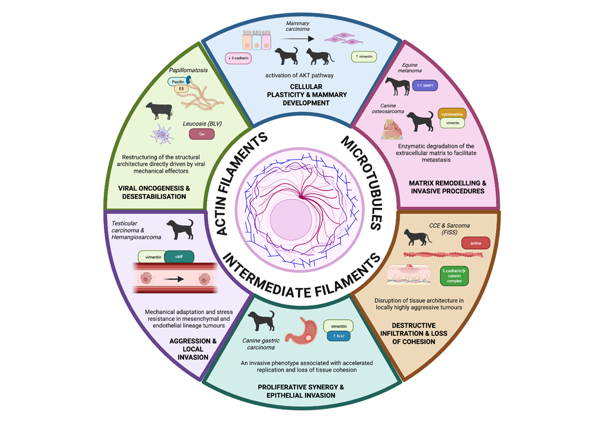
Figure 2: Integrated schematic of cytoskeletal remodeling associated with tumor aggressiveness in veterinary oncology. The central diagram shows the interaction between actin filaments, microtubules, and intermediate filaments as a coordinated functional network. The surrounding panels present species- and tumor-specific contexts in which cytoskeletal reorganization is associated with malignant behavior. Actin dynamics contribute to cellular plasticity and the formation of invasive structures, particularly in mammary carcinoma and other epithelial neoplasms. Microtubules participate in proliferative signaling and intracellular organization, including processes associated with AKT pathway activation. Intermediate filaments, especially vimentin, contribute to mechanical adaptation, resistance to stress, and epithelial-mesenchymal plasticity. Additional panels include extracellular matrix remodeling mediated by metalloproteinases in equine melanoma, cytoskeletal alterations associated with viral oncoproteins in bovine papillomavirus-associated neoplasia and enzootic bovine leukosis, and disruption of tissue cohesion linked to the E-cadherin/β-catenin complex in aggressive feline carcinomas. The figure integrates these alterations across species and relates them to invasion, metastasis, and tumor progression. Created with BioRender.com.
Comparison with human medicine
From the perspective of the cytoskeleton as a functional axis of tumor aggressiveness, comparisons with human medicine are particularly relevant. Comparative oncology integrates findings from veterinary and human medicine and recognizes that several fundamental mechanisms of carcinogenesis are shared among mammals (Sultan & Ganaie, 2018). Unlike experimental models of induced cancer in rodents, companion animals develop tumors spontaneously in immunocompetent organisms and under environmental exposures shared with humans. These characteristics allow spontaneous tumors in dogs and cats to recapitulate relevant aspects of the heterogeneity and biological complexity of human cancer, supporting their value in comparative oncology (Gardner et al., 2016; Oh & Cho, 2023).
One of the most relevant examples of this comparative approach is mammary carcinoma. Canine and feline mammary tumors share several histopathological and biological characteristics with human breast cancer, including certain patterns of lymphatic and hematogenous dissemination. Abdelmegeed and Mohammed (2018) highlighted that alterations associated with epithelial-mesenchymal transition (EMT) exhibit conserved features across species. In tumors of epithelial origin, decreased E-cadherin expression and aberrant vimentin expression may reflect epithelial-mesenchymal plasticity and have been associated with invasive behavior. These similarities support the value of spontaneous mammary tumors, particularly canine mammary tumors, as comparative models for investigating mechanisms associated with cytoskeletal remodeling and tumor invasion (Abdelmegeed & Mohammed, 2018).
Genomic studies have further strengthened this perspective by identifying alterations in cancer-associated genes and signaling pathways shared between canine and human tumors (Bird & Smith, 2025). These molecular similarities complement the histopathological and biological parallels observed across species and support the comparative investigation of mechanisms associated with tumor progression. From a cytoskeletal perspective, pathways involved in cell adhesion, migration, adaptation to stress, and microtubule dynamics are particularly relevant, and some of their components are targets of antineoplastic agents used in both human and veterinary oncology (Oh & Cho, 2023).
Finally, the study of spontaneous tumors in companion animals offers complementary advantages over conventional experimental models because these tumors develop in the presence of an intact immune system and exhibit intratumoral heterogeneity and natural interactions with the tumor microenvironment. Therefore, investigating cytoskeletal remodeling and its association with tumor-cell migration, invasion, and adaptation to the microenvironment in dogs and cats may provide translationally relevant information for human oncology (Bird & Smith, 2025; Sultan & Ganaie, 2018). Within this context, comparative veterinary oncology provides a valuable framework for identifying conserved mechanisms of tumor progression and exploring potential therapeutic targets relevant to both animal and human health.
Future perspectives
The study of the cytoskeleton in veterinary oncology represents a field with still underexplored diagnostic, prognostic, and therapeutic potential. Proteins such as vimentin are used as markers of cellular lineage or differentiation, whereas adhesion molecules such as E-cadherin provide information on the maintenance or loss of the epithelial phenotype. Beyond their utility as markers, available evidence indicates that the reorganization of these structural and adhesion networks contributes to tumor-cell responses to mechanical and metabolic stress within the microenvironment (Danielsson et al., 2018). Characterization of these alterations may contribute to the identification of novel prognostic biomarkers and potential therapeutic targets related to cytoskeletal dynamics (Ritter & Kreis, 2022).
An important challenge for future research will be to elucidate the role of post-translational modifications (PTMs), such as phosphorylation and glycosylation, in regulating cytoskeletal proteins and their functions across different cellular and tumor contexts (Parvanian & Eriksson, 2025; Satelli & Li, 2011). In addition, the identification of cytoskeleton-associated proteins in extracellular vesicles raises the possibility of exploring liquid-biopsy approaches in veterinary oncology. However, their utility as biomarkers of tumor aggressiveness or metastatic progression requires specific validation in veterinary patients and across different tumor types (Parvanian & Eriksson, 2025).
From an experimental perspective, conventional two-dimensional cultures can be complemented by three-dimensional systems and organoids derived from animal tumors. These models provide more representative platforms for investigating mechanotransduction, cytoskeletal remodeling, and cellular responses to tumor microenvironmental conditions, including hypoxia and extracellular matrix stiffness (Nosalova et al., 2024). Because spontaneous tumors in animals share several molecular and biological characteristics with their human counterparts, incorporating these models may strengthen their translational value within comparative oncology (Oh & Cho, 2023). Furthermore, approaches such as spatial transcriptomics and single-cell sequencing could help identify tumor-cell subpopulations and hybrid states of cellular plasticity that cannot be fully characterized using conventional immunohistochemistry (Bird & Smith, 2025; Parvanian & Eriksson, 2025; Sahoo et al., 2022).
Finally, this review highlights the need to expand research to species and tumor types for which evidence regarding the cytoskeleton remains limited, such as uterine adenocarcinoma in rabbits and insulinoma in ferrets. In these cases, species- and tumor-specific evidence will be necessary to determine whether cytoskeletal alterations are associated with particular stages of neoplastic progression, invasive behavior, or clinical prognosis, rather than relying exclusively on extrapolations from human oncology. Investigating the cytoskeleton as a dynamic functional network may contribute to a more precise understanding of the mechanisms underlying tumor progression and, potentially, to the identification of new diagnostic, prognostic, and therapeutic strategies in veterinary oncology.
Conclusion
The cytoskeleton constitutes an integrative functional axis that connects biochemical and molecular signals with cellular responses to the mechanical properties of the tumor microenvironment. Based on the evidence reviewed in dogs, cats, horses, and cattle, cytoskeletal remodeling is recurrently associated with cellular plasticity, invasion, and other features of tumor aggressiveness, although the strength and mechanistic basis of these associations vary among tumor types and species.
Within this process, actin reorganization plays a central role in cell migration and invasion, whereas aberrant vimentin expression in neoplastic cells of epithelial origin and the loss or altered localization of E-cadherin have been associated with epithelial-mesenchymal plasticity and, in several tumor types, with more aggressive biological behavior. Likewise, microtubule dynamics and intracellular transport contribute to processes essential for tumor-cell proliferation, polarity, and survival. However, the interpretation of these alterations must consider cellular lineage and neoplastic context, distinguishing the physiological expression of cytoskeletal proteins from their aberrant expression, overexpression, or reorganization during tumor progression.
Nevertheless, future research in veterinary oncology should more rigorously distinguish immunohistochemical and clinicopathological associations from functional mechanisms directly involved in invasion and metastasis. Although the available evidence supports an association between cytoskeletal remodeling and tumor aggressiveness, functional validation of these processes in veterinary neoplasms remains limited for some tumor types, and part of their mechanistic interpretation is still derived from experimental models or human oncology.
Within the framework of comparative oncology, the study of spontaneous tumors in domestic animals reinforces their value as translational biological models. Understanding the cytoskeleton not merely as a structural framework but as a dynamic network involved in cellular plasticity, mechanical adaptation, and invasive behavior may contribute to refining diagnostic and prognostic strategies and identifying potential therapeutic targets relevant to both veterinary and human oncology.
Acknowledgments
The authors acknowledge the Facultad de Estudios Superiores Cuautitlán (FESC), Campus 4, Universidad Nacional Autónoma de México, for the facilities and academic support provided during the development of this work. This research was supported by the PAPIME project PE214226.
References
Abdelmegeed, S., & Mohammed, S. (2018). Canine mammary tumors as a model for human disease (Review). Oncology Letters, 15(6), 8195-8205. https://doi.org/10.3892/ol.2018.8411
Akhmanova, A., & Steinmetz, M. O. (2015). Control of microtubule organization and dynamics: Two ends in the limelight. Nature Reviews Molecular Cell Biology, 16(12), 711-726. https://doi.org/10.1038/nrm4084
Amaral, C. B., Leite, J. da S., Fonseca, A. B. M., & Ferreira, A. M. R. (2018). Vimentin, osteocalcin and osteonectin expression in canine primary bone tumors: Diagnostic and prognostic implications. Molecular Biology Reports, 45(5), 1289-1296. https://doi.org/10.1007/s11033-018-4285-6
Armando, F., Godizzi, F., Razzuoli, E., Leonardi, F., Angelone, M., Corradi, A., Meloni, D., Ferrari, L., & Passeri, B. (2020). Epithelial to mesenchymal transition (EMT) in a laryngeal squamous cell carcinoma of a horse: Future perspectives. Animals, 10(12), 2318. https://doi.org/10.3390/ani10122318
Asproni, P., Millanta, F., Ressel, L., Podestà, F., Parisi, F., Vannozzi, I., & Poli, A. (2021). An immunohistochemical study of the PTEN/AKT pathway involvement in canine and feline mammary tumors. Animals, 11(2), 365. https://doi.org/10.3390/ani11020365
Bird, R. C., & Smith, B. F. (2025). Comparative genetics of canine and human cancers. Veterinary Sciences, 12(9), 875. https://doi.org/10.3390/vetsci12090875
Borzacchiello, G., & Roperto, F. (2008). Bovine papillomaviruses, papillomas and cancer in cattle. Veterinary Research, 39(5), 45. https://doi.org/10.1051/vetres:2008022
Bravo-Cordero, J. J., Hodgson, L., & Condeelis, J. (2012). Directed cell invasion and migration during metastasis. Current Opinion in Cell Biology, 24(2), 277-283. https://doi.org/10.1016/j.ceb.2011.12.004
Brodesser, D. M., Kummer, S., Eichberger, J. A., Schlangen, K., Corteggio, A., Borzacchiello, G., Bertram, C. A., Brandt, S., & Pratscher, B. (2024). Deregulation of metalloproteinase expression in gray horse melanoma ex vivo and in vitro. Cells, 13(11), 956. https://doi.org/10.3390/cells13110956
Caliari, D., Zappulli, V., Rasotto, R., Cardazzo, B., Frassineti, F., Goldschmidt, M. H., & Castagnaro, M. (2014). Triple-negative vimentin-positive heterogeneous feline mammary carcinomas as a potential comparative model for breast cancer. BMC Veterinary Research, 10(1), 185. https://doi.org/10.1186/s12917-014-0185-8
Danielsson, F., Peterson, M. K., Caldeira Araújo, H., Lautenschläger, F., & Gad, A. K. B. (2018). Vimentin diversity in health and disease. Cells, 7(10), 147. https://doi.org/10.3390/cells7100147
de Barros, M. D., Dos Santos Batista, B. P., Amaral, C. B., da Silva Leite, J., & Ferreira, A. M. R. (2025). Vimentin expression in canine testicular tumors: A local invasion predictive biomarker. Biotechnic & Histochemistry, 100(4), 208-215. https://doi.org/10.1080/10520295.2025.2492397
de Cecco, B. S., Argenta, F. F., Bianchi, R. M., De Lorenzo, C., Wronski, J. G., Bandinelli, M. B., da Costa, F. V., Driemeier, D., Pavarini, S. P., & Sonne, L. (2021). Feline giant-cell pleomorphic sarcoma: Cytologic, histologic and immunohistochemical characterization. Journal of Feline Medicine and Surgery, 23(8), 738-744. https://doi.org/10.1177/1098612X20972667
Fife, C. M., McCarroll, J. A., & Kavallaris, M. (2014). Movers and shakers: Cell cytoskeleton in cancer metastasis. British Journal of Pharmacology, 171(24), 5507-5523. https://doi.org/10.1111/bph.12704
Flores, A. R., Rêma, A., Mesquita, J. R., Taulescu, M., Seixas, F., Gärtner, F., & Amorim, I. (2022). Vimentin and Ki-67 immunolabeling in canine gastric carcinomas and their prognostic value. Veterinary Pathology, 59(6), 903-914. https://doi.org/10.1177/03009858221117858
Gamba, C. O., Rodrigues, M. A., Gomes, D. A., Estrela-Lima, A., Ferreira, E., & Cassali, G. D. (2015). The relationship between E-cadherin and its transcriptional repressors in spontaneously arising canine invasive micropapillary mammary carcinoma. Journal of Comparative Pathology, 153(4), 256-265. https://doi.org/10.1016/j.jcpa.2015.08.006
Gardner, H. L., Fenger, J. M., & London, C. A. (2016). Dogs as a model for cancer. Annual Review of Animal Biosciences, 4(1), 199-222. https://doi.org/10.1146/annurev-animal-022114-110911
Gillet, N., Florins, A., Boxus, M., Burteau, C., Nigro, A., Vandermeers, F., Balon, H., Bouzar, A. B., Defoiche, J., Burny, A., Reichert, M., Kettmann, R., & Willems, L. (2007). Mechanisms of leukemogenesis induced by bovine leukemia virus: Prospects for novel anti-retroviral therapies in human. Retrovirology, 4, 18. https://doi.org/10.1186/1742-4690-4-18
Hanahan, D. (2022). Hallmarks of cancer: New dimensions. Cancer Discovery, 12(1), 31-46. https://doi.org/10.1158/2159-8290.CD-21-1059
Hardas, A., Suárez-Bonnet, A., Beck, S., Becker, W. E., Ramírez, G. A., & Priestnall, S. L. (2021). Canine gastric carcinomas: A histopathological and immunohistochemical study and similarities with the human counterpart. Animals, 11(5), 1409. https://doi.org/10.3390/ani11051409
Harris, K., Gelberg, H. B., Kiupel, M., & Helfand, S. C. (2019). Immunohistochemical features of epithelial-mesenchymal transition in feline oral squamous cell carcinoma. Veterinary Pathology, 56(6), 826-839. https://doi.org/10.1177/0300985819859873
Hartmann, K., Day, M. J., Thiry, E., Lloret, A., Frymus, T., Addie, D., Boucraut-Baralon, C., Egberink, H., Gruffydd-Jones, T., Horzinek, M. C., Hosie, M. J., Lutz, H., Marsilio, F., Pennisi, M. G., Radford, A. D., Truyen, U., & Möstl, K. (2015). Feline injection-site sarcoma: ABCD guidelines on prevention and management. Journal of Feline Medicine and Surgery, 17(7), 606-613. https://doi.org/10.1177/1098612X15588451
Herrmann, H., & Aebi, U. (2016). Intermediate filaments: Structure and assembly. Cold Spring Harbor Perspectives in Biology, 8(11), a018242. https://doi.org/10.1101/cshperspect.a018242
Lane, A. (2018). Tumors in domestic animals. Australian Veterinary Journal, 96(12), 507. https://doi.org/10.1111/avj.12624
Li, X., & Wang, J. (2020). Mechanical tumor microenvironment and transduction: Cytoskeleton mediates cancer cell invasion and metastasis. International Journal of Biological Sciences, 16(12), 2014-2028. https://doi.org/10.7150/ijbs.44943
Maniscalco, L., Iussich, S., Martín de las Mulas, J., Millán, Y., Biolatti, B., Sasaki, N., Nakagawa, T., & De Maria, R. (2012). Activation of AKT in feline mammary carcinoma: A new prognostic factor for feline mammary tumours. The Veterinary Journal, 191(1), 65-71. https://doi.org/10.1016/j.tvjl.2010.12.016
Martinez Cuesta, L., Nieto Farias, M. V., Lendez, P. A., Rowland, R. R. R., Sheahan, M. A., Cheuquepán Valenzuela, F. A., Marin, M. S., Dolcini, G., & Ceriani, M. C. (2019). Effect of bovine leukemia virus on bovine mammary epithelial cells. Virus Research, 271, 197678. https://doi.org/10.1016/j.virusres.2019.197678
Mendez, M. G., Kojima, S., & Goldman, R. D. (2010). Vimentin induces changes in cell shape, motility, and adhesion during the epithelial to mesenchymal transition. The FASEB Journal, 24(6), 1838-1851. https://doi.org/10.1096/fj.09-151639
Nagamine, E., Hirayama, K., Matsuda, K., Okamoto, M., Ohmachi, T., Kadosawa, T., & Taniyama, H. (2015). Diversity of histologic patterns and expression of cytoskeletal proteins in canine skeletal osteosarcoma. Veterinary Pathology, 52(5), 977-984. https://doi.org/10.1177/0300985815574006
Nosalova, N., Huniadi, M., Horňáková, Ľ., Valenčáková, A., Horňák, S., Nagoos, K., Vozar, J., & Cizkova, D. (2024). Canine mammary tumors: Classification, biomarkers, traditional and personalized therapies. International Journal of Molecular Sciences, 25(5), 2891. https://doi.org/10.3390/ijms25052891
Oh, J. H., & Cho, J.-Y. (2023). Comparative oncology: Overcoming human cancer through companion animal studies. Experimental & Molecular Medicine, 55(4), 725-734. https://doi.org/10.1038/s12276-023-00977-3
Ong, M. S., Deng, S., Halim, C. E., Cai, W., Tan, T. Z., Huang, R. Y.-J., Sethi, G., Hooi, S. C., Kumar, A. P., & Yap, C. T. (2020). Cytoskeletal proteins in cancer and intracellular stress: A therapeutic perspective. Cancers, 12(1), 238. https://doi.org/10.3390/cancers12010238
Paoloni, M. C., & Khanna, C. (2007). Comparative oncology today. Veterinary Clinics of North America: Small Animal Practice, 37(6), 1023-1032. https://doi.org/10.1016/j.cvsm.2007.08.003
Parvanian, S., & Eriksson, J. E. (2025). Vimentin in the tumor microenvironment: Orchestrating invasion, immunity, and metabolism. European Journal of Cell Biology, 104(4), 151523. https://doi.org/10.1016/j.ejcb.2025.151523
Pimenta, J., Pires, I., Prada, J., & Cotovio, M. (2023). E-cadherin immunostaining in equine melanocytic tumors. Animals, 13(13), 2216. https://doi.org/10.3390/ani13132216
Ritter, A., & Kreis, N.-N. (2022). Microtubule dynamics and cancer. Cancers, 14(18), 4368. https://doi.org/10.3390/cancers14184368
Sabattini, S., & Bettini, G. (2009). An immunohistochemical analysis of canine haemangioma and haemangiosarcoma. Journal of Comparative Pathology, 140(2-3), 158-168. https://doi.org/10.1016/j.jcpa.2008.10.006
Sahoo, S., Ashraf, B., Duddu, A. S., Biddle, A., & Jolly, M. K. (2022). Interconnected high-dimensional landscapes of epithelial-mesenchymal plasticity and stemness in cancer. Clinical & Experimental Metastasis, 39(2), 279-290. https://doi.org/10.1007/s10585-021-10139-2
Sammarco, A., Gomiero, C., Beffagna, G., Cavicchioli, L., Ferro, S., Michieletto, S., Orvieto, E., Patruno, M., & Zappulli, V. (2023). Epithelial-to-mesenchymal transition and phenotypic marker evaluation in human, canine, and feline mammary gland tumors. Animals, 13(5), 878. https://doi.org/10.3390/ani13050878
Sanz Ressel, B. L., Gomez Castro, G., Massone, A. R., & Barbeito, C. G. (2023). Altered E-cadherin/β-catenin expression in feline cutaneous squamous cell carcinomas. Journal of Comparative Pathology, 207, 21-24. https://doi.org/10.1016/j.jcpa.2023.09.007
Satelli, A., & Li, S. (2011). Vimentin in cancer and its potential as a molecular target for cancer therapy. Cellular and Molecular Life Sciences, 68(18), 3033-3046. https://doi.org/10.1007/s00018-011-0735-1
Strohmayer, C., Klang, A., Kummer, S., Walter, I., Jindra, C., Weissenbacher-Lang, C., Redmer, T., Kneissl, S., & Brandt, S. (2022). Tumor cell plasticity in equine papillomavirus-positive versus-negative squamous cell carcinoma of the head and neck. Pathogens, 11(2), 266. https://doi.org/10.3390/pathogens11020266
Sultan, F., & Ganaie, B. A. (2018). Comparative oncology: Integrating human and veterinary medicine. Open Veterinary Journal, 8(1), 25. https://doi.org/10.4314/ovj.v8i1.5
Tsirimonaki, E., Ullah, R., Marchetti, B., Ashrafi, G. H., McGarry, L., Ozanne, B., & Campo, M. S. (2006). Similarities and differences between the E5 oncoproteins of bovine papillomaviruses type 1 and type 4: Cytoskeleton, motility and invasiveness in E5-transformed bovine and mouse cells. Virus Research, 115(2), 158-168. https://doi.org/10.1016/j.virusres.2005.08.003
Usman, S., Waseem, N. H., Nguyen, T. K. N., Mohsin, S., Jamal, A., Teh, M.-T., & Waseem, A. (2021). Vimentin is at the heart of epithelial mesenchymal transition (EMT) mediated metastasis. Cancers, 13(19), 4985. https://doi.org/10.3390/cancers13194985
Vail, D. M., Thamm, D. H., & Liptak, J. M. (2020). Withrow & MacEwen’s small animal clinical oncology (6th ed.). Saunders.
Vogelstein, B., Papadopoulos, N., Velculescu, V. E., Zhou, S., Diaz, L. A., & Kinzler, K. W. (2013). Cancer genome landscapes. Science, 339(6127), 1546-1558. https://doi.org/10.1126/science.1235122
Yamaguchi, H., & Condeelis, J. (2007). Regulation of the actin cytoskeleton in cancer cell migration and invasion. Biochimica et Biophysica Acta (BBA) - Molecular Cell Research, 1773(5), 642-652. https://doi.org/10.1016/j.bbamcr.2006.07.001
Zappulli, V., Rasotto, R., Caliari, D., Mainenti, M., Peña, L., Goldschmidt, M. H., & Kiupel, M. (2015). Prognostic evaluation of feline mammary carcinomas: A review of the literature. Veterinary Pathology, 52(1), 46-60. https://doi.org/10.1177/0300985814528221
Conflict of interest statement
The authors declare no conflicts of interest.
Ethical considerations
Not applicable. This study was a literature review and did not involve experimental procedures in animals or humans.
Author contributions
1. Conceptualization, 2. Investigation, 3. Supervision, 4. Writing - original draft, 5. Writing - review & editing.
Sarai Guerrero-López contributed to 1, 2, and 4. Carlos Gerardo García-Tovar to 3 and 5. Samantha Jardon-Xicotencatl to 1, 3 and 5. All authors read and approved the final version of the manuscript.
Nota del editor
El editor José Manuel Verdes aprobó este artículo